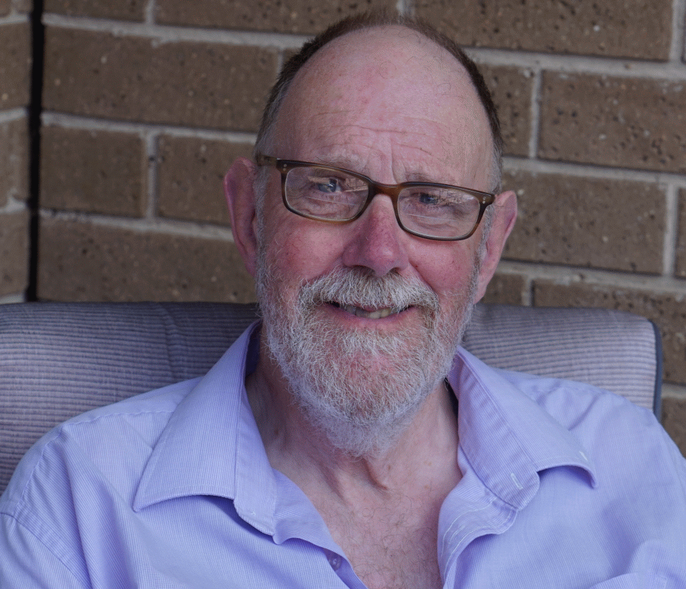
Neville Curtis
PhD in Organic Chemistry from the University of Alberta 1981
Research Fellow at the Australian National University from 1981-84
Research Scientist at the Defence Science and Technology Organisation (DSTO) from 1984-2014
Visiting Professor at the Australian Defence Force Academy @ the University of New South Wales
Over 130 reviewed publications in:
· Organic Chemistry
· Coordination Chemistry
· Energetic materials
· Soft Operations Research
Fellow of the Operational Research Society (UK)
Received NATO Science and Technology prize 2013
new opal discussions
Neville offers some new insights and discussion for the analysis of opal by X-Ray Diffraction and offers mineralogical information. You can read more here:
I am a relative newcomer to the field of opals. After retiring from DSTO, Professor Allan Pring (then at the South Australian Museum and Flinders University) interested me in looking at whether the original Jones and Segnit X-ray diffraction-based classification of opals into opal-AG, opal-AN, opal-CT and opal-C still worked with all the recent discoveries being made in new locations. As it turned out, it did, at least as a primary tool, but I found plenty of chemistry-based questions to excite my interest. In particular, I wasn’t convinced that the classification was complete or that opal-CT was a homogenous group. I also thought that the chemist’s bag of tricks hadn’t been adequately exploited and that more could be learned by widening the scope of characterisation. Perhaps a chemist’s approach would complement that of mineralogists and geologists. Between golf rounds I put in about 2 days a week.
Amongst things that gave me a “feeling of unease” (a Soft Operations Research term) was the notion of a diagenesis of opal-A through opal-CT and opal-C to quartz/moganite. This appeared to me to be informed by observation not experiment and based on the notion that older samples (e.g. around hot springs) appeared to be opal-CT and younger ones, opal-A. From my experience with aging of nitrocellulose in gun propellants I am well aware of the perils of artificial accelerated aging and making bold predictions without a time machine. While it may be right, it is not demonstrated. I was also unconvinced that the x-ray diffraction patterns of opal-CT could be explained in terms of a mix of cristobalite and tridymite. In contrast, opal-C looks to be wet, impure cristobalite.
Finally (of course) I was interested in whether I could crack the structure of the various forms of opal and perhaps provide insights into how opal is formed.
I was thus able to bring my general knowledge and skills in chemical characterisation and kinetics, along with access to a wide range of worldwide samples to look at opals. For the past few years I’ve been working with associate Professor Martin Johnston and Dr Jason Gascooke at Flinders University (the Flinders Opal Group, or FOG for short) taking a systematic approach to the use of various techniques while at the same time making sure we have enough samples in the study to avoid being misled by a rogue opal. We’ve achieved grants to use the facilities at the Australian Synchroton and the Taipan neutron line at Lucas Heights. The South Australian Museum has supported me through a Research Associateship while I am an adjunct Professor at Flinders University. I am grateful for this access to samples and scientific instruments. Through presentation at the 2021 Coober Pedy National Opal Symposium I’ve made contact with some of the other players in the field. So far, we’ve looked at over 250 different samples using x-ray diffraction, Raman and infrared vibrational spectroscopy, 1H and 29Si nuclear magnetic resonance spectroscopy, inelastic neutron scattering, neutron diffraction and scanning electron microscopy. I’m extending the initial study by looking at opals from many worldwide sites. Two things that I’ve noticed are: there are not that many worldwide opal-AG sites (play of colour may also be opal-CT) and that opal-C samples are few and far between. My feeling is that our repository of analyses would be a valuable resource to the isolated opaleers scattered around the globe.
I’m happy to correspond with anyone with a question or observation about opals.
A presentation by Neville Curtis at the opal symposium 2021
A review of the classification of opal with reference to recent new localities, N.J. Curtis, J.R. Gascooke, M.R. Johnston and A. Pring (2019) Minerals paper 299: You can read the full article here:
Abstract: Our examination of over 230 worldwide opal samples shows that X-ray diffraction (XRD) remains the best primary method for delineation and classification of opal-A, opal-CT and opal-C, though we found that mid-range infra-red spectroscopy provides an acceptable alternative. Raman, infra-red and nuclear magnetic resonance spectroscopy may also provide additional information to assist in classification and provenance. The corpus of results indicated that the opal-CT group covers a range of structural states and will benefit from further multi-technique analysis. At the one end are the opal-CTs that provide a simple XRD pattern (“simple” opal-CT) that includes Ethiopian play-of-colour samples, which are not opal-A. At the other end of the range are those opal-CTs that give a complex XRD pattern (“complex” opal-CT). The majority of opal-CT samples fall at this end of the range, though some show play-of-colour. Raman spectra provide some correlation. Specimens from new opal finds were examined. Those from Ethiopia, Kazakhstan, Madagascar, Peru, Tanzania and Turkey all proved to be opal-CT. Of the three specimens examined from Indonesian localities, one proved to be opal-A, while a second sample and the play-of-colour opal from West Java was a “simple” Opal-CT. Evidence for two transitional types having characteristics of opal-A and opal-CT, and “simple” opal-CT and opal-C are presented.
Silicon-oxygen region infrared and Raman analysis of opals: the effect of sample preparation and measurement type, N.J. Curtis, J.R. Gascooke and A. Pring (2021) Minerals paper 173. You can read the full paper here:
Abstract: An extensive infrared (IR) spectroscopy study using transmission, specular and diffuse reflectance, and attenuated total reflection (ATR) was undertaken to characterise opal-AG, opal- AN (hyalite), opal-CT and opal-C, focusing on the Si-O fingerprint region (200–1600 cm−1). We show that IR spectroscopy is a viable alternative to X-ray powder diffraction (XRD) as a primary means of classification of opals even when minor levels of impurities are present. Variable angle specular reflectance spectroscopy shows that the three major IR bands of opal are split into transverse optical (TO) and longitudinal optical (LO) components. Previously observed variability in powder ATR is probably linked to the very high refractive index of opals at infrared wavelengths, rather than heterogeneity or particle size effects. An alternative use of ATR using unpowdered samples provides a potential means of non-destructive delineation of play of colour opals into opal-AG or opal-CT gems. We find that there are no special structural features in the infrared spectrum that differentiate opal from silica glasses. Evidence is presented that suggests silanol environments may be responsible for the structural differences between opal-AG, opal-AN and other forms of opaline silica. Complementary studies with Raman spectroscopy, XRD and scanning electron microscopy (SEM) provide evidence of structural trends within the opal-CT type.
29Si Solid-State NMR Analysis of Opal-AG, Opal-AN and Opal-CT: Single Pulse Spectroscopy and Spin-Lattice T1 Relaxometry, N.J. Curtis, J.R. Gascooke, M.R. Johnston and A. Pring (2022) Minerals paper 323. You can read the full text here:
Abstract: Single pulse, solid-state 29Si nuclear magnetic resonance (NMR) spectroscopy offers an additional method of characterisation of opal-A and opal-CT through spin-lattice (T1) relaxometry. Opal T1 relaxation is characterised by stretched exponential (Weibull) function represented by scale (speed of relaxation) and shape (form of the curve) parameters. Relaxation is at least an order of magnitude faster than for silica glass and quartz, with Q3 (silanol) usually faster than Q4 (fully substituted silicates). 95% relaxation (Q4) is achieved for some Australian seam opals after 50 s though other samples of opal-AG may take 4000 s, while some figures for opal-AN are over 10,000 s. Enhancement is probably mostly due to the presence of water/silanol though the presence of paramagnetic metal ions and molecular motion may also contribute. Shape factors for opal-AG (0.5) and opal-AN (0.7) are significantly different, consistent with varying water and silanol environments, possibly reflecting differences in formation conditions. Opal-CT samples show a trend of shape factors from 0.45 to 0.75 correlated to relaxation rate. Peak position, scale and shape parameter, and Q3 to Q4 ratios offer further differentiating feature to separate opal-AG and opal-AN from other forms of opaline silica. T1 relaxation measurement may have a role for provenance verification. In addition, definitively determined Q3/Q4 ratios are in the range 0.1 to 0.4 for opal-AG but considerably lower for opal-AN and opal-CT.


